A graduate-level optical-engineering study of single-photon microscopy, presented with a Python classical simulation of quantum-dot emission demonstrating reconstruction resolution beyond the natural (diffraction) limit.
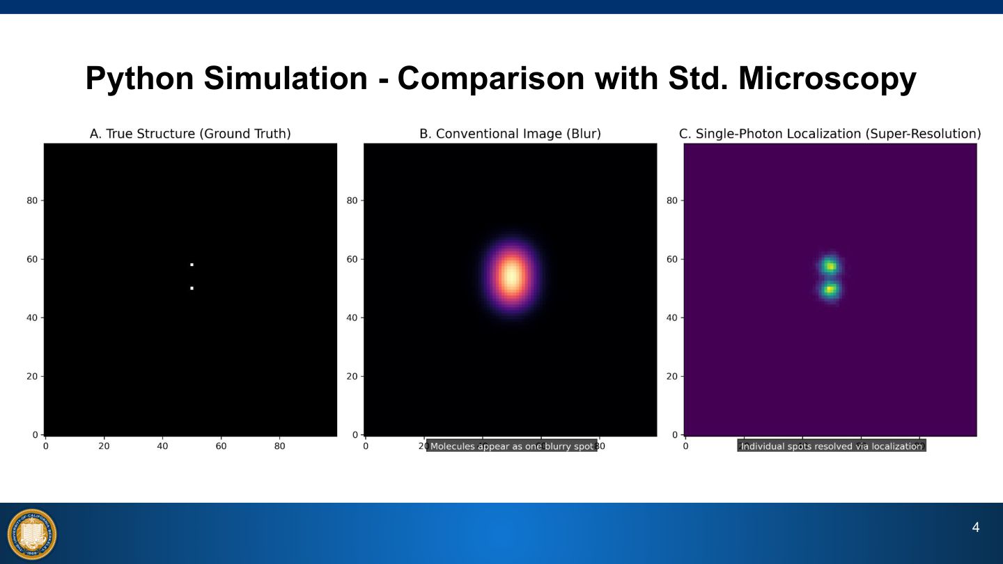
Single-photon microscopy uses quantum emitters embedded in a specimen (quantum dots, organic dyes, and fluorescent proteins) to isolate individual point-spread functions, enabling localization precision that breaks the classical diffraction resolution limit.
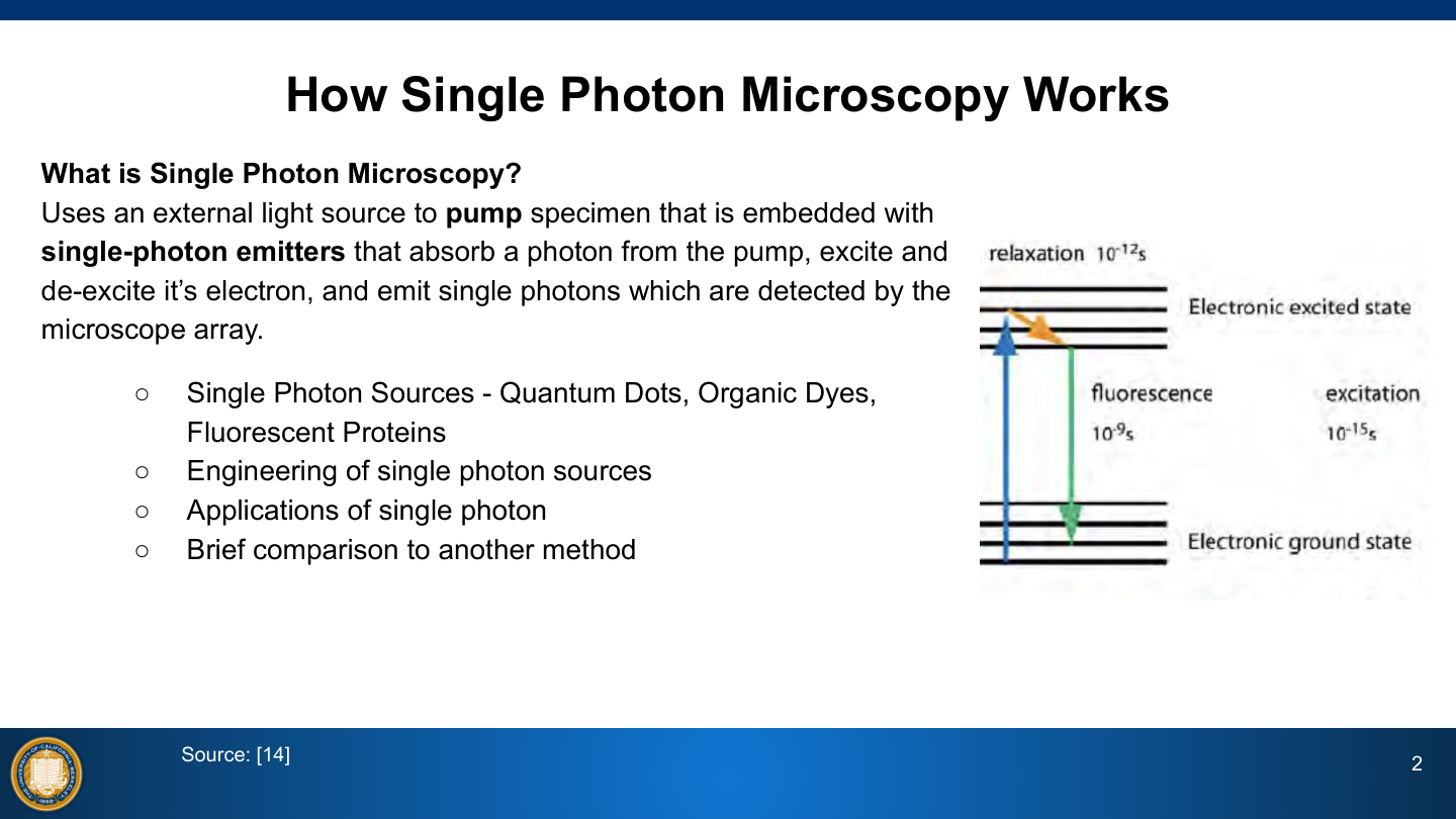
The three classes of single-photon source each involve distinct tradeoffs: quantum dots offer high photostability but exhibit stochastic blinking; organic dyes achieve smaller linkage errors but require cytotoxic imaging buffers; fluorescent proteins are biocompatible and genetically encoded but have low photon counts and poor photostability.
Spontaneous emission from these sources is governed by quantum electrodynamics via Dirac radiation theory, with key parameters, molar extinction coefficient, fluorescence quantum yield, and fluorescence lifetime, determining the practical imaging performance of each emitter type.
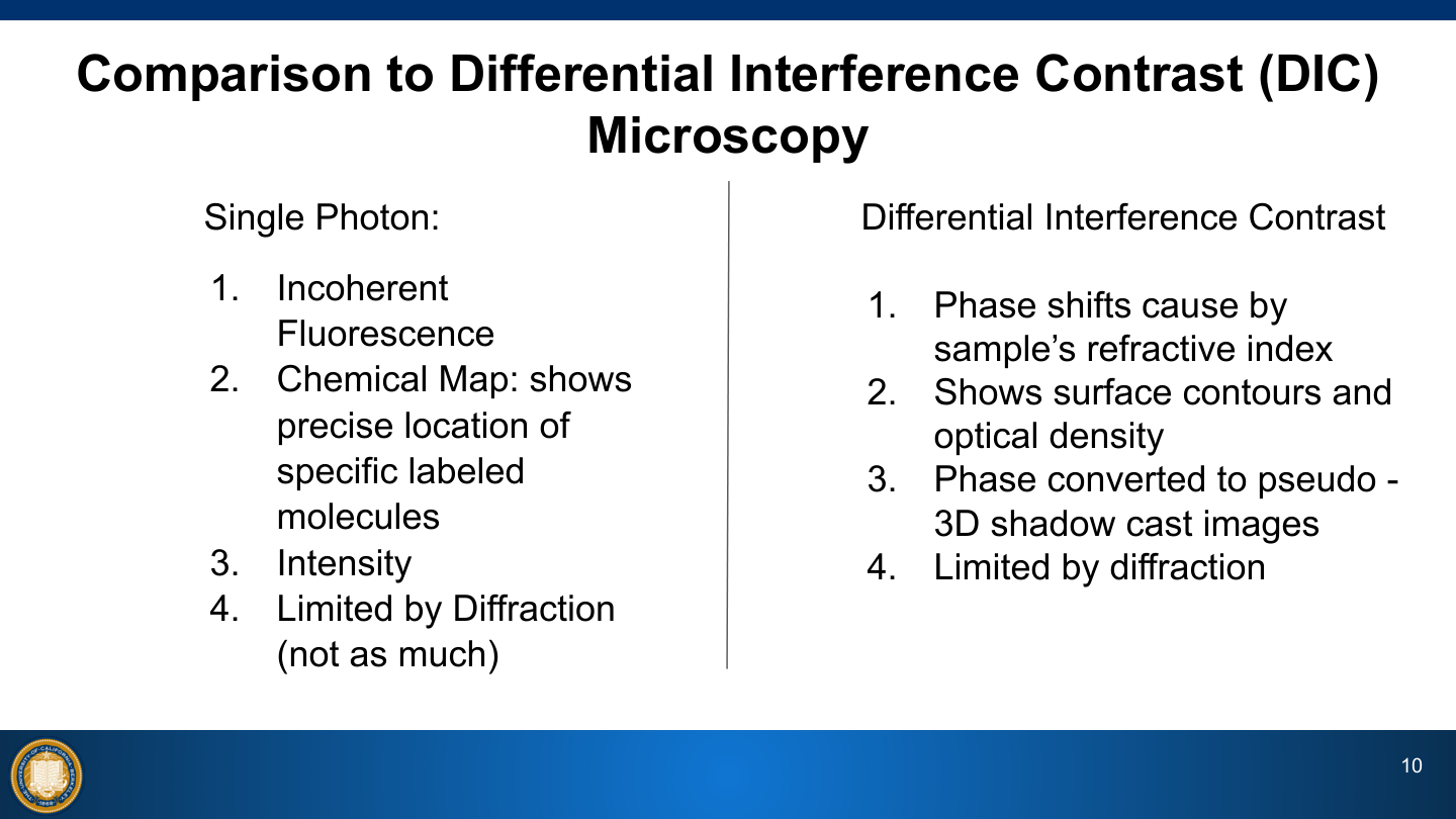
Compared to Differential Interference Contrast microscopy, single-photon methods produce chemical maps of specific labeled-molecule locations via incoherent fluorescence rather than phase-derived surface-contour images, offering complementary structural information at reduced diffraction-limited constraints.